Mammograms explained: A mammogram is the Medical way of finding and demonstrating the human breast. Mammography is a staged event and may comprise the following: -
The first step is to have the X-ray based mammogram. Here, the breast is examined in a special machine designed for obtaining excellent images of the breast. Four pictures are taken of the breasts, each one taken in two directions. Processing of the images can be either analogue which are film based or digital which is all electronic. There is no difference in the accuracy or diagnostic efficiency of either method. The breast must be gently compressed and never ever hurt during the examination because it is soft and requires to be still for the picture taking and also the compression improves the image quality. In a modern machine, the compression is computer controlled and is released themoment the exposure is completed. there are very strict criteria established for high quality mammography and this examination must only be performed by highly skilled and specially trained technicians and specialist doctors.
Mammograms save lives and it has been proven that by having annual mammograms, the death rate for breast cancer will decrease by 38%. If the time period is extended to every two years, the death rate will be doubled.
It is now recommended that every woman should have an annual mammogram therefore from the age of 40 years and because we are finding more and more young women getting breast cancer some people are advising women to start at 35 years.
In excess of 90% of examinations it will be necessary to complete a mammogram with an Ultrasound study. Here again, high definition dedicated apparatus must be used by a highly skilled operator and in SA this will invariably be a Doctor and not a technician.
The reason for using ultrasound is because certain things can be seen with X-ray based technology and others only with Ultrasound. Each examination on its own therefore, is incomplete for mammography and would miss many breast disease. The two together, however, complete the examination and result in almost 100% accuracy. it is very important to realize that an X-ray mammogram on its own, whether analogue or digital is an incomplete study and should not be done in the modern context of accurate diagnosis.
In a small percentage of cases, if the above cannot make an exact diagnosis, the next stage would be a Breast Biopsy. |
||
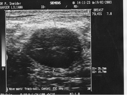
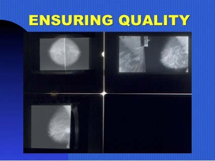 TOP........QUALITY MAMMOGRAMS ARE VERY IMPORTANT, TOP LEFT; TUMOUR MISSED AS SHOWN BOTTOM WHEN PROPERLY TAKEN. TOP RIGHT TUMOUR OF RIGHT MAMMOGRAM MISSED AS SHOWN IN THE LEFT HAND PICTURE. BOTTOM.......ULTRASOUND ILLUSTRATION OF A BENIGN BREAST TUMOUR. |
 |
|